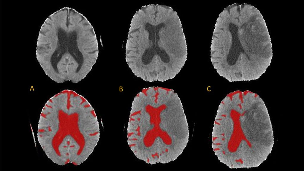
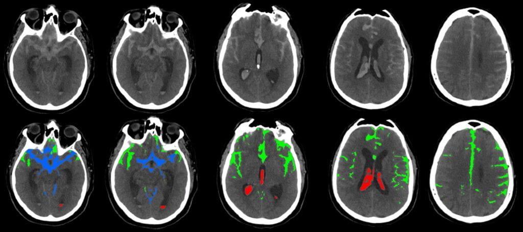
Our lab seeks to leverage data and imaging-driven approaches to understand the heterogeneity of human responses to severe brain injuries. It is our vision that all patients with acute brain injuries, from ischemic stroke to hemorrhage and brain trauma, will have their care precisely tailored to their own individual clinical and biologic status.
With generous support …
Our lab operates with support from the National Institutes of Neurological Disorders and Stroke under grants U24-NS-132940 and R01-NS-121218.
We also receive support from the Mid-America Transplant Foundation and the Alvin Siteman Cerebrovascular Research Fund.
News
Long-standing hormone treatment for donated hearts found to be ineffective (Links to an external site)
Practice of using thyroid hormones to preserve function for transplantation may even cause harm


